Gelatin/PLGA Microspheres as a 3D Scaffold for Chondrocytes
DOI:
https://doi.org/10.54133/ajms.v6i2.898Keywords:
Chondrocytes, Gelatin, Microspheres, PLGAAbstract
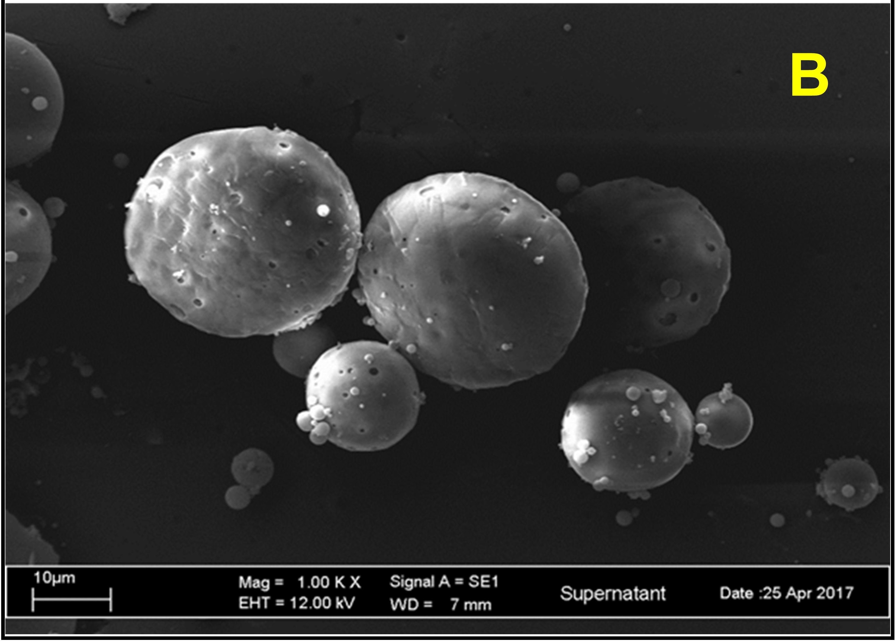
Background: Osteoarthritis (OA) degrades cartilage and bone. Osteochondral autograft, allograft, and total replacement knee surgery have limitations, such as prompt immune responses, lack of cartilage tissue obtainability, invasiveness, and a loosening implant that may require further correction. Tissue engineering, which involves injecting chondrocytes into 3D porous scaffold carriers in the joint, seems promising for tissue repair and growth. Objective: To develop gelatin/poly DL-lactide-co-glycolide (PLGA) microspheres as a porous scaffold for chondrocyte carriers. Methods: The double emulsion method is one of the most popular and best methods for forming microspheres. In summary, in the PLGA oil phase, we emulsified a gelatin solution representing the inner aqueous phase. Next, in an external aqueous phase of polyvinyl alcohol (PVA), we emulsified the resultant first emulsion. The double emulsion was stirred to evaporate organic solvent and centrifuged to collect gelatin and PLGA microspheres. Results: The Mastersizer result showed polydispersed particles with 23.53% of the desirable cell injection size range between 1-300 µm. Scanning electronic microscope (SEM) images revealed spherical and porous microspheres with smooth surfaces. The average absolute zeta potential value was -30.7±4.895, indicating stable preparation. Conclusions: Gelatin and PLAGA polymers worked together to make 3D scaffold microspheres that were the right size, had the right number of holes, and were strong.
Downloads
References
Yao Q, Wu X, Tao C, Gong W, Chen M, Qu M, et al. Osteoarthritis: pathogenic signaling pathways and therapeutic targets. Signal Transduct Target Ther. 2023;8:56. doi: 10.1038/s41392-023-01330-w.
da Costa BR, Reichenbach S, Keller N, Nartey L, Wandel S, Jüni P, et al. Effectiveness of non-steroidal anti-inflammatory drugs for the treatment of pain in knee and hip osteoarthritis: a network meta-analysis. Lancet. 2017;390(10090):e21–33. doi: 10.1016/S0140-6736(17)31744-0.
Ayhan E, Kesmezacar H, Akgun I. Intraarticular injections (corticosteroid, hyaluronic acid, platelet rich plasma) for the knee osteoarthritis. World J Orthop. 2014;5(3):351–361. doi: 10.5312/wjo.v5.i3.351.
Montoya F, Martínez F, García-Robles M, Balmaceda-Aguilera C, Koch X, Rodríguez F, et al. Clinical and experimental approaches to knee cartilage lesion repair and mesenchymal stem cell chondrocyte differentiation. Biol Res. 2013;46(4):441–451. doi: 10.4067/S0716-97602013000400015.
Parel PM, Manyak GA, Carvajal JA, Abraham T, Al Rashid M. An early-stage comparison of functional outcomes following robotic-assisted versus conventional total knee arthroplasty: a systematic review and meta-analysis. J Arthrosc Jt Surg. 2022;9(3):77. doi: 10.4103/jajs.jajs_75_22.
Zhang L, Hu J, Athanasiou KA. The role of tissue engineering in articular cartilage repair and regeneration. Crit Rev Biomed Engneer. 2009;37(1–2):1–57. doi: 10.1615/critrevbiomedeng.v37.i1-2.10.
Solouk A, Mirzadeh H, Amanpour S. Injectable scaffold as minimally invasive technique for cartilage tissue engineering: in vitro and in vivo preliminary study. Prog Biomater. 2014;3:143–151. doi: 10.1007/s40204-014-0031-x.
Zhang Z. Injectable biomaterials for stem cell delivery and tissue regeneration. Expert Opin Biol Ther. 2017;17(1):49–62. doi: 10.1080/14712598.2017.1256389.
Kadam NR. Microsphere: a brief review. Asian J Biomed Pharm Sci. 2015;5(47):13-19. doi: 10.15272/ajbps.v5i47.713.
Malda J, van Blitterswijk CA, Grojec M, Martens DE, Tramper J, Riesle J. Expansion of bovine chondrocytes on microcarriers enhances redifferentiation. Tissue Eng. 2003;9(5):939–948. doi: 10.1089/107632703322495583.
Hossain KMZ, Patel U, Ahmed I. Development of microspheres for biomedical applications: a review. Prog Biomater. 2015;4(1):1–19. doi: 10.1007/s40204-014-0033-8.
Zhao C, Zhu Z, Cao X, Pan F, Li F, Xue M, et al. Evaluation the injectability of injectable microparticle delivery systems on the basis of injection force and discharged rate. Eur J Pharm Biopharm. 2023;190:58–72. doi: 10.1016/j.ejpb.2023.06.017.
Elzoghby AO. Gelatin-based nanoparticles as drug and gene delivery systems: reviewing three decades of research. J Control Release. 2013;172(3):1075–1091. doi: 10.1016/j.jconrel.2013.09.019.
Makadia HK, Siegel SJ. Poly cactic-co-glycolic acid (PLGA) as biodegradable controlled drug delivery carrier. Polymers (Basel). 2011;3(3):1377–1397. doi: 10.3390/polym3031377.
Yang H, Yao L, Wang Y, Chen G, Chen H. Advancing cell surface modification in mammalian cells with synthetic molecules. Chem Sci. 2023;14(46):13325–13345. doi: 10.1039/D3SC04597H.
Vedadghavami A, He T, Zhang C, Amiji SM, Hakim B, Bajpayee AG. Charge-based drug delivery to cartilage: Hydrophobic and not electrostatic interactions are the dominant cause of competitive binding of cationic carriers in synovial fluid. Acta Biomater. 2022;151:278–289. doi: 10.1016/j.actbio.2022.08.010.
Tsung MJ, Burgess DJ. Preparation and characterization of gelatin surface modified PLGA microspheres. AAPS PharmSci. 2001;3(2):E11. doi: 10.1208/ps030211.
Yasuda T. Cartilage destruction by matrix degradation products. Mod Rheumatol. 2006;16(4):197–205. doi: 10.1007/s10165-006-0490-6.
Chun KW, Yoo HS, Yoon JJ, Park TG. Biodegradable PLGA microcarriers for injectable delivery of chondrocytes: effect of surface modification on cell attachment and function. Biotechnol Prog. 2004;20(6):1797–1801. doi: 10.1021/bp0496981.
Mehrasa M, Asadollahi MA, Ghaedi K, Salehi H, Arpanaei A. Electrospun aligned PLGA and PLGA/gelatin nanofibers embedded with silica nanoparticles for tissue engineering. Int J Biol Macromol. 2015;79:687–695. doi: 10.1016/j.ijbiomac.2015.05.050.
Banerjee I, Mishra D, Maiti TK. PLGA microspheres incorporated gelatin scaffold: microspheres modulate scaffold properties. Int J Biomater. 2009;2009:143659. doi: 10.1155/2009/143659.
Tan H, Huang D, Lao L, Gao C. RGD modified PLGA/gelatin microspheres as microcarriers for chondrocyte delivery. J Biomed Mater Res B Appl Biomater. 2009;91(1):228–238. doi: 10.1002/jbm.b.31394.
Jain RA. The manufacturing techniques of various drug loaded biodegradable poly(lactide-co-glycolide) (PLGA) devices. Biomater. 2000;21(23):2475–2490. doi: 10.1016/S0142-9612(00)00115-0.
Morhenn VB, Lemperle G, Gallo RL. Phagocytosis of different particulate dermal filler substances by human macrophages and skin cells. Dermatol Surg. 2002;28(6):484–490. doi: 10.1046/j.1524-4725.2002.01273.x.
Zilberman M, Grinberg O. HRP-loaded bioresorbable microspheres: effect of copolymer composition and molecular weight on microstructure and release profile. J Biomater Appl. 2008; 22(5):391-407. doi: 10.1177/0885328207077591.
Choudhury M, Mohanty S, Nayak S. Effect of different solvents in solvent casting of porous pla scaffolds—in biomedical and tissue engineering applications. J Tissue Sci Eng. 2015;5(1):1–9. doi: 10.4172/2157-7552.1000142.
Pandya N, Pandya M, Bhaskar VH. Preparation and in vitro characterizatin of porous carrier–based glipizide floating microspheres for gastric delivery. J Young Pharm. 2011;3(2):97–104. doi: 10.4103/0975-1483.80292.
Ghanbar H, Luo CJ, Bakhshi P, Day R, Edirisinghe M. Preparation of porous microsphere-scaffolds by electrohydrodynamic forming and thermally induced phase separation. Mater Sci Eng C. 2013;33(5):2488–2498. doi: 10.1016/j.msec.2012.12.098.
Manaia EB, Abuçafy MP, Chiari-Andréo BG, Silva BL, Oshiro Junior JA, Chiavacci LA. Physicochemical characterization of drug nanocarriers. Int J Nanomed. 2017;12:4991–5011. doi: 10.2147/IJN.S133832.
Ruan G, Feng SS. Preparation and characterization of poly (lactic acid)-poly(ethylene glycol)-poly(lactic acid) (PLA-PEG-PLA) microspheres for controlled release of paclitaxel. Biomater. 2003;24(27):5037–5044. doi: 10.1016/s0142-9612(03)00419-8.
Guo C, Gemeinhart RA. Understanding the adsorption mechanism of chitosan onto poly(lactide-co-glycolide) particles. Eur J Pharm Biopharm. 2008;70(2):597–604. doi: 10.1016/j.ejpb.2008.06.008.
Downloads
Published
How to Cite
Issue
Section
License
Copyright (c) 2024 Al-Rafidain Journal of Medical Sciences ( ISSN 2789-3219 )

This work is licensed under a Creative Commons Attribution-NonCommercial-ShareAlike 4.0 International License.
Published by Al-Rafidain University College. This is an open access journal issued under the CC BY-NC-SA 4.0 license (https://creativecommons.org/licenses/by-nc-sa/4.0/).


