Gelatin/PLGA Microspheres as a 3D Scaffold for Chondrocytes
DOI:
https://doi.org/10.54133/ajms.v6i2.898الكلمات المفتاحية:
الكرات المجهرية، جيلاتين، بولي (حمض اللاكتيك المشارك الجليكوليك)، الخلايا الغضروفيةالملخص
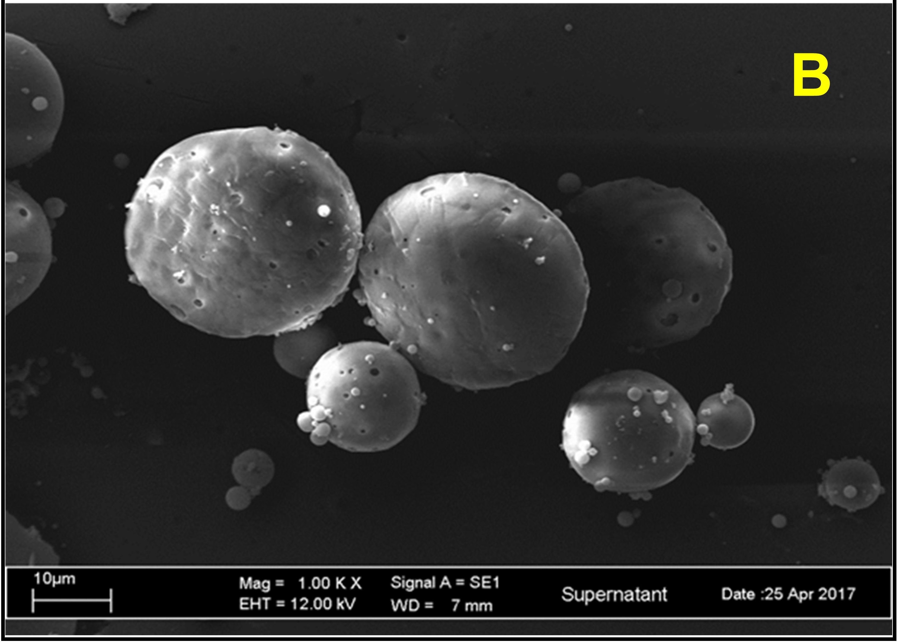
هشاشة العظام (OA) هو مرض المفاصل الذي ينطوي على تدهور أنسجة الغضاريف والبنية العظمية الأساسية. تُظهر الأساليب الثلاثة التي تم تطبيقها لإصلاح الغضروف التالف، مثل الطعم الذاتي والطعم الخيفي وجراحة الاستبدال الكلي، قيودًا بسبب الاستجابات المناعية السريعة، وعدم إمكانية الحصول على أنسجة الغضروف، والتداخل الجراحي، وفقدان الغضروف المستبدل التي قد تحتاج إلى المزيد جراحة التصحيح. توفر هندسة الأنسجة استراتيجية واعدة لإصلاح وتجديد الأنسجة التالفة من خلال دمجها في حاملات ناقلة مسامية ثلاثية الأبعاد متبوعة بزرعها باستخدام جهاز قابل للحقن. لذلك، قد تم تطوير الكرات المجهرية الجيلاتينية/PLGA لتكون بمثابة نقالة مسامية لحامل الخلايا الغضروفية. تعد طريقة المستحلب المزدوج إحدى الطرق الشائعة وأفضلها المستخدمة لتكوين الكرات المجهرية. باختصار، تم استحلاب محلول الجيلاتين الذي يمثل الطور المائي الداخلي في مرحلة زيت PLGA. ثم تم استحلاب المستحلب الأول الناتج في الطور المائي الخارجي من PVA. تم تحريك المستحلب المزدوج لتبخير المذيب العضوي وطرده مركزيًا لتجميع كريات الجيلاتين/PLGA المجهرية. أظهرت نتيجة Mastersizer أن جزيئات متعددة التشتت مع 23.53% من حجم حقن الخلايا المرغوب فيه الذي يتراوح بين 1-300 مايكرومتر. تكشف صور SEM عن كرة مجهرية كروية ومسامية ذات أسطح ناعمة. وكان متوسط قيمة إمكانات زيتا المطلقة -30.7±4.895، مما يدل على استقرار المستحضر. في الختام، كانت بوليمرات الجيلاتين/PLGA قادرة على العمل بشكل تآزري وصياغة كريات مجهرية ثلاثية الأبعاد بالحجم المرغوب والمسامية والقوة الميكانيكية
التنزيلات
المراجع
Yao Q, Wu X, Tao C, Gong W, Chen M, Qu M, et al. Osteoarthritis: pathogenic signaling pathways and therapeutic targets. Signal Transduct Target Ther. 2023;8:56. doi: 10.1038/s41392-023-01330-w.
da Costa BR, Reichenbach S, Keller N, Nartey L, Wandel S, Jüni P, et al. Effectiveness of non-steroidal anti-inflammatory drugs for the treatment of pain in knee and hip osteoarthritis: a network meta-analysis. Lancet. 2017;390(10090):e21–33. doi: 10.1016/S0140-6736(17)31744-0.
Ayhan E, Kesmezacar H, Akgun I. Intraarticular injections (corticosteroid, hyaluronic acid, platelet rich plasma) for the knee osteoarthritis. World J Orthop. 2014;5(3):351–361. doi: 10.5312/wjo.v5.i3.351.
Montoya F, Martínez F, García-Robles M, Balmaceda-Aguilera C, Koch X, Rodríguez F, et al. Clinical and experimental approaches to knee cartilage lesion repair and mesenchymal stem cell chondrocyte differentiation. Biol Res. 2013;46(4):441–451. doi: 10.4067/S0716-97602013000400015.
Parel PM, Manyak GA, Carvajal JA, Abraham T, Al Rashid M. An early-stage comparison of functional outcomes following robotic-assisted versus conventional total knee arthroplasty: a systematic review and meta-analysis. J Arthrosc Jt Surg. 2022;9(3):77. doi: 10.4103/jajs.jajs_75_22.
Zhang L, Hu J, Athanasiou KA. The role of tissue engineering in articular cartilage repair and regeneration. Crit Rev Biomed Engneer. 2009;37(1–2):1–57. doi: 10.1615/critrevbiomedeng.v37.i1-2.10.
Solouk A, Mirzadeh H, Amanpour S. Injectable scaffold as minimally invasive technique for cartilage tissue engineering: in vitro and in vivo preliminary study. Prog Biomater. 2014;3:143–151. doi: 10.1007/s40204-014-0031-x.
Zhang Z. Injectable biomaterials for stem cell delivery and tissue regeneration. Expert Opin Biol Ther. 2017;17(1):49–62. doi: 10.1080/14712598.2017.1256389.
Kadam NR. Microsphere: a brief review. Asian J Biomed Pharm Sci. 2015;5(47):13-19. doi: 10.15272/ajbps.v5i47.713.
Malda J, van Blitterswijk CA, Grojec M, Martens DE, Tramper J, Riesle J. Expansion of bovine chondrocytes on microcarriers enhances redifferentiation. Tissue Eng. 2003;9(5):939–948. doi: 10.1089/107632703322495583.
Hossain KMZ, Patel U, Ahmed I. Development of microspheres for biomedical applications: a review. Prog Biomater. 2015;4(1):1–19. doi: 10.1007/s40204-014-0033-8.
Zhao C, Zhu Z, Cao X, Pan F, Li F, Xue M, et al. Evaluation the injectability of injectable microparticle delivery systems on the basis of injection force and discharged rate. Eur J Pharm Biopharm. 2023;190:58–72. doi: 10.1016/j.ejpb.2023.06.017.
Elzoghby AO. Gelatin-based nanoparticles as drug and gene delivery systems: reviewing three decades of research. J Control Release. 2013;172(3):1075–1091. doi: 10.1016/j.jconrel.2013.09.019.
Makadia HK, Siegel SJ. Poly cactic-co-glycolic acid (PLGA) as biodegradable controlled drug delivery carrier. Polymers (Basel). 2011;3(3):1377–1397. doi: 10.3390/polym3031377.
Yang H, Yao L, Wang Y, Chen G, Chen H. Advancing cell surface modification in mammalian cells with synthetic molecules. Chem Sci. 2023;14(46):13325–13345. doi: 10.1039/D3SC04597H.
Vedadghavami A, He T, Zhang C, Amiji SM, Hakim B, Bajpayee AG. Charge-based drug delivery to cartilage: Hydrophobic and not electrostatic interactions are the dominant cause of competitive binding of cationic carriers in synovial fluid. Acta Biomater. 2022;151:278–289. doi: 10.1016/j.actbio.2022.08.010.
Tsung MJ, Burgess DJ. Preparation and characterization of gelatin surface modified PLGA microspheres. AAPS PharmSci. 2001;3(2):E11. doi: 10.1208/ps030211.
Yasuda T. Cartilage destruction by matrix degradation products. Mod Rheumatol. 2006;16(4):197–205. doi: 10.1007/s10165-006-0490-6.
Chun KW, Yoo HS, Yoon JJ, Park TG. Biodegradable PLGA microcarriers for injectable delivery of chondrocytes: effect of surface modification on cell attachment and function. Biotechnol Prog. 2004;20(6):1797–1801. doi: 10.1021/bp0496981.
Mehrasa M, Asadollahi MA, Ghaedi K, Salehi H, Arpanaei A. Electrospun aligned PLGA and PLGA/gelatin nanofibers embedded with silica nanoparticles for tissue engineering. Int J Biol Macromol. 2015;79:687–695. doi: 10.1016/j.ijbiomac.2015.05.050.
Banerjee I, Mishra D, Maiti TK. PLGA microspheres incorporated gelatin scaffold: microspheres modulate scaffold properties. Int J Biomater. 2009;2009:143659. doi: 10.1155/2009/143659.
Tan H, Huang D, Lao L, Gao C. RGD modified PLGA/gelatin microspheres as microcarriers for chondrocyte delivery. J Biomed Mater Res B Appl Biomater. 2009;91(1):228–238. doi: 10.1002/jbm.b.31394.
Jain RA. The manufacturing techniques of various drug loaded biodegradable poly(lactide-co-glycolide) (PLGA) devices. Biomater. 2000;21(23):2475–2490. doi: 10.1016/S0142-9612(00)00115-0.
Morhenn VB, Lemperle G, Gallo RL. Phagocytosis of different particulate dermal filler substances by human macrophages and skin cells. Dermatol Surg. 2002;28(6):484–490. doi: 10.1046/j.1524-4725.2002.01273.x.
Zilberman M, Grinberg O. HRP-loaded bioresorbable microspheres: effect of copolymer composition and molecular weight on microstructure and release profile. J Biomater Appl. 2008; 22(5):391-407. doi: 10.1177/0885328207077591.
Choudhury M, Mohanty S, Nayak S. Effect of different solvents in solvent casting of porous pla scaffolds—in biomedical and tissue engineering applications. J Tissue Sci Eng. 2015;5(1):1–9. doi: 10.4172/2157-7552.1000142.
Pandya N, Pandya M, Bhaskar VH. Preparation and in vitro characterizatin of porous carrier–based glipizide floating microspheres for gastric delivery. J Young Pharm. 2011;3(2):97–104. doi: 10.4103/0975-1483.80292.
Ghanbar H, Luo CJ, Bakhshi P, Day R, Edirisinghe M. Preparation of porous microsphere-scaffolds by electrohydrodynamic forming and thermally induced phase separation. Mater Sci Eng C. 2013;33(5):2488–2498. doi: 10.1016/j.msec.2012.12.098.
Manaia EB, Abuçafy MP, Chiari-Andréo BG, Silva BL, Oshiro Junior JA, Chiavacci LA. Physicochemical characterization of drug nanocarriers. Int J Nanomed. 2017;12:4991–5011. doi: 10.2147/IJN.S133832.
Ruan G, Feng SS. Preparation and characterization of poly (lactic acid)-poly(ethylene glycol)-poly(lactic acid) (PLA-PEG-PLA) microspheres for controlled release of paclitaxel. Biomater. 2003;24(27):5037–5044. doi: 10.1016/s0142-9612(03)00419-8.
Guo C, Gemeinhart RA. Understanding the adsorption mechanism of chitosan onto poly(lactide-co-glycolide) particles. Eur J Pharm Biopharm. 2008;70(2):597–604. doi: 10.1016/j.ejpb.2008.06.008.
التنزيلات
منشور
كيفية الاقتباس
إصدار
القسم
الرخصة
الحقوق الفكرية (c) 2024 Al-Rafidain Journal of Medical Sciences

هذا العمل مرخص بموجب Creative Commons Attribution-NonCommercial-ShareAlike 4.0 International License.
Published by Al-Rafidain University College. This is an open access journal issued under the CC BY-NC-SA 4.0 license (https://creativecommons.org/licenses/by-nc-sa/4.0/).