A Comparative in vitro Investigation on the Impact of Fiber Insertion on Cuspal Deflection of Maxillary Premolars Restored with Various Types of Bulk Fill Direct Composite Restorations
DOI:
https://doi.org/10.54133/ajms.v5i.179Keywords:
Bulk fill composite restorations, Cuspal deflection, Direct posterior restoration, Fiber reinforcement composite, Glass fiberAbstract
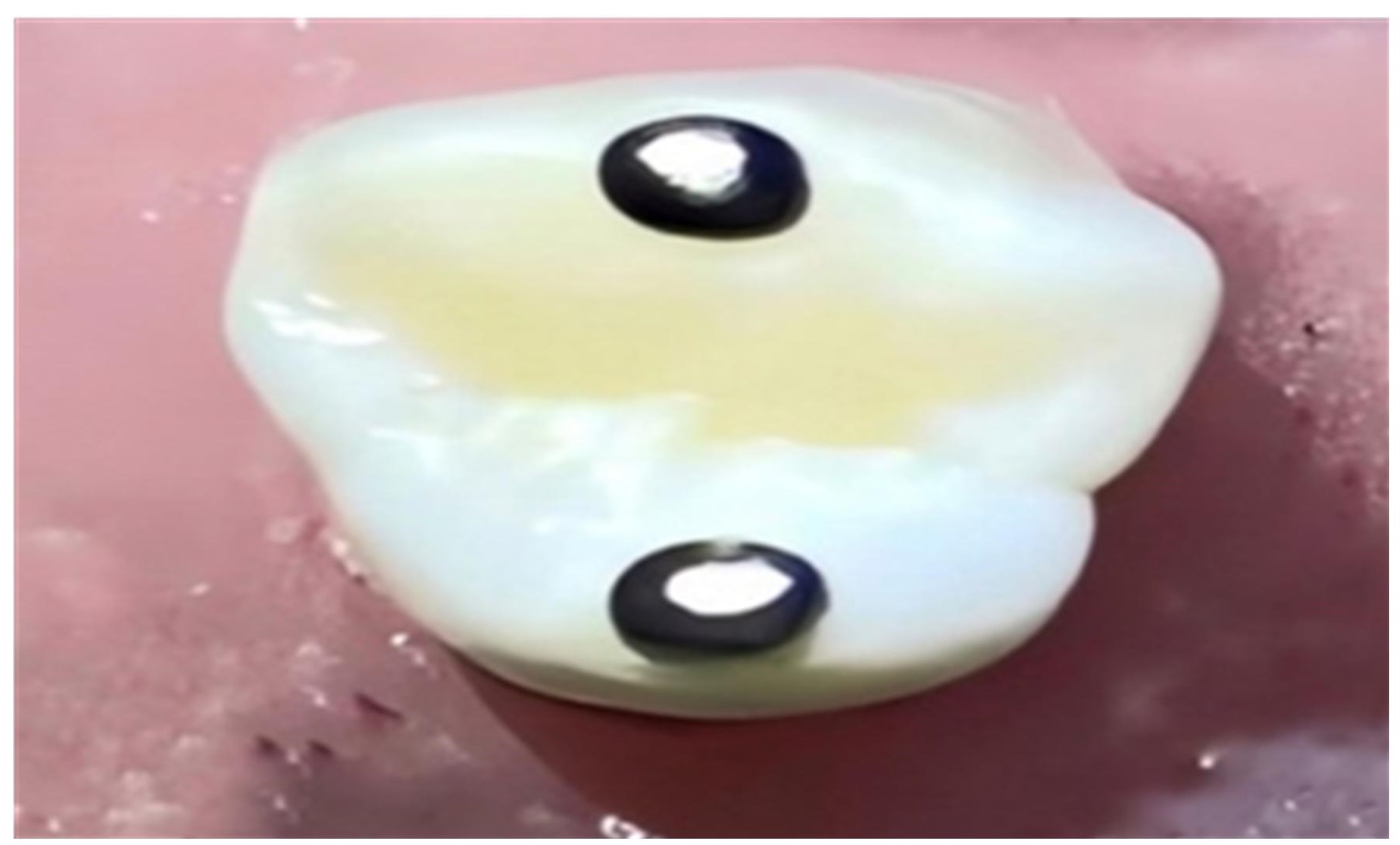
Background: Polymerization shrinkage remains a significant disadvantage that makes the use of direct composite restorations difficult. Objective: To evaluate how well a fiber insert and multiple bulk-fill direct restorations worked on the cuspal deflection of MOD cavities in premolar teeth using a Dino-Lite digital microscope and computer software. Methods: In sixty fresh maxillary first premolars, large MOD cavities were created. Teeth were randomly divided into six groups of ten based on restorative materials. SonicFill®, Beautifil Bulk®, and Filtek® Bulk Fill posterior restoratives were used in groups A1, B1, and C1, whereas groups A2, B2, and C2 used the same bulk composite with E-glass fiber (UFM, Dentapreg). Under a digital microscope, the intercuspal distance between two reference points on the cusp tips was measured before preparation, after preparation, 15 minutes after finishing restorations, and one month following incubation. Results: There was a significant difference between the groups after 15 minutes of restoration, but no significant differences following cavity preparation or one month of incubation. CD values were considerably higher after 15 minutes of restoration in groups restored with bulk fill only. Beautifil Bulk Fill restorative resulted in greater cuspal deflection than the other groups. The CD values in each group were significantly higher 15 minutes after restoration compared to cavity preparation and a month of incubation. Conclusion: Using inserts, cuspal deflection in MOD cavities can be significantly minimized, and stress release usually occurs after water incubation.
Downloads
References
Demirel G, Baltacioglu I, Kolsuz M, Ocak M, Bilecenoglu B, Orhan K. Volumetric cuspal deflection of premolars restored with different paste-like bulk-fill resin composites evaluated by microcomputed tomography. Oper Dent. 2020;45(2):143-50. doi: 10.2341/19-019-L. DOI: https://doi.org/10.2341/19-019-L
Dilian NS, Kadhim AJ. Comparative evaluation of marginal microleakage between bulk-fill, preheated Bulk-Fill, and Bulk-Fill flowable composite resins above and below cemento-enamel junction using micro-computed tomography: An in vitro study. Dent Hypotheses. 2022;13(4):128. doi: 10.4103/denthyp.denthyp_104_22. DOI: https://doi.org/10.4103/denthyp.denthyp_104_22
Jafarpour S, El-Badrawy W, Jazi HS, McComb D. Effect of composite insertion technique on cuspal deflection using an in vitro simulation model. Oper Dent. 2012;37(3):299-305. doi: 10.2341/11-086-L. DOI: https://doi.org/10.2341/11-086-L
Elsharkasi M, Platt J, Cook N, Yassen G, Matis B. Cuspal deflection in premolar teeth restored with bulk-fill resin-based composite materials. Oper Dent. 2018;43(1):E1-E9. doi: 10.2341/16-072-L. DOI: https://doi.org/10.2341/16-072-L
Karaman E, Ozgunaltay G. Cuspal deflection in premolar teeth restored using current composite resins with and without resin-modified glass ionomer liner. Oper Dent. 2013;38(3):282-9. doi: 10.2341/11-400-L. DOI: https://doi.org/10.2341/11-400-L
Yaroub M, Hameed MR. Evaluation of marginal gap at the composite/enamel interface in class II composite resin restoration by SEM after thermal and mechanical load cycling: An in vitro comparative study. J Baghdad Coll Dent. 2014;325(2217):1-8. DOI: https://doi.org/10.12816/0015256
Kim M, Park S. Comparison of premolar cuspal deflection in bulk or in incremental composite restoration methods. Oper Dent. 2011;36(3):326-334. doi: 10.2341/10-315-L. DOI: https://doi.org/10.2341/10-315-L
Safwat EM, Khater AG, Abd-Elsatar AG, Khater GA. Glass fiber-reinforced composites in dentistry. Bull Natl Res Cent. 2021;45:1-9. doi: 10.1186/s42269-021-00650-7. DOI: https://doi.org/10.1186/s42269-021-00650-7
Ibrahim BH, Al-Azzawi HJ. Fracture resistance of endodontically treated premolar teeth with extensive MOD cavities restored with different Bulk Fill composite restorations (An in vitro study). J. Baghdad Coll Dent. 2017;29(2):26-32. doi: 10.12816/0038746. DOI: https://doi.org/10.12816/0038746
Chieruzzi M, Pagano S, Pennacchi M, Lombardo G, D’Errico P, Kenny JM. Compressive and flexural behaviour of fibre reinforced endodontic posts. J Dent. 2012;40(11):968-978. doi: 10.1016/j.jdent.2012.08.003. DOI: https://doi.org/10.1016/j.jdent.2012.08.003
Campos EA, Andrade MF, Porto-Neto ST, Campos LA, Saad JRC, Deliberador TM, et al. Cuspal movement related to different bonding techniques using etch-and-rinse and self-etch adhesive systems. Eur J Dent. 2009;3(03):213-218. doi: 10.1016/j.jdent.2012.08.003. DOI: https://doi.org/10.1055/s-0039-1697434
Dhingra V, Taneja S, Kumar M, Kumari M. Influence of fiber inserts, type of composite, and gingival margin location on the microleakage in Class II resin composite restorations. Oper Dent. 2014;39(1):E9-E15. doi: 10.2341/12-349-L. DOI: https://doi.org/10.2341/12-349-L
Jlekh ZA, Abdul-Ameer ZM. Evaluation of the cuspal deflection of premolars restored with different types of bulk fill composite restoration. Biomed Pharmacol J. 2018;11(2):751-757. doi: 10.13005/bpj/1429. DOI: https://doi.org/10.13005/bpj/1429
Salem HN, Elhefnawy SH, Moharam LM. Effect of different restoration techniques and cavity designs on cuspal deflection of posterior teeth restored with resin composite inlays. Futur Dent J. 2018;4(2):146-149. doi: 10.1016/j.fdj.2018.09.004. DOI: https://doi.org/10.1016/j.fdj.2018.09.004
González López S, Sanz Chinesta MV, Ceballos García L, de Haro Gasquet F, González Rodríguez MP. Influence of cavity type and size of composite restorations on cuspal flexure. Med Oral Patol Oral Cir Bucal. 2006;11(6):E536-540.
Shabayek N, Hassan F, Mobarak E. Effect of using silorane-based resin composite for restoring conservative cavities on the changes in cuspal deflection. Oper Dent. 2013;38(2):E42-E9. doi: 10.2341/12-035-L. DOI: https://doi.org/10.2341/12-035-L
Behery H, El‐Mowafy O, El‐Badrawy W, Saleh B, Nabih S. Cuspal deflection of premolars restored with bulk‐fill composite resins. J Esthet Resto Dent. 2016;28(2):122-30. doi: 10.1111/jerd.12188. DOI: https://doi.org/10.1111/jerd.12188
Francis A, Braxton A, Ahmad W, Tantbirojn D, Simon J, Versluis A. Cuspal flexure and extent of cure of a bulk-fill flowable base composite. Oper Dent. 2015;40(5):515-523. doi: 10.2341/14-235-L. DOI: https://doi.org/10.2341/14-235-L
Tsujimoto A, Nagura Y, Barkmeier WW, Watanabe H, Johnson WW, Takamizawa T, et al. Simulated cuspal deflection and flexural properties of high viscosity bulk-fill and conventional resin composites. J. Mech. Behav. Biomed. Mater. 2018;87:111-118. doi: 10.1016/j.jmbbm.2018.07.013. DOI: https://doi.org/10.1016/j.jmbbm.2018.07.013
Versluis A, Tantbirojn D, Lee MS, Tu LS, DeLong R. Can hygroscopic expansion compensate polymerization shrinkage? Part I. Deformation of restored teeth. Dent Mater. 2011;27(2):126-133. 10.1016/j.dental.2010.09.007. DOI: https://doi.org/10.1016/j.dental.2010.09.007
Blažić L, Pantelić D, Savić-Šević S, Murić B, Belić I, Panić B. Modulated photoactivation of composite restoration: measurement of cuspal movement using holographic interferometry. Lasers Med Sci. 2011;26:179-186. doi: 10.1007/s10103-010-0774-0. DOI: https://doi.org/10.1007/s10103-010-0774-0
Ólafsson VG, Ritter AV, Swift EJ, Boushell LW, Ko CC, Jackson GR, et al. Effect of composite type and placement technique on cuspal strain. J Esthet Resto Dent. 2018;30(1):30-38. doi: 10.1111/jerd.12339. DOI: https://doi.org/10.1111/jerd.12339
Singhal S, Gurtu A, Singhal A, Bansal R, Mohan S. Effect of different composite restorations on the cuspal deflection of premolars restored with different insertion techniques-an in vitro study. J Clin Diagnostic Res. 2017;11(8):ZC67. doi: 10.7860/jcdr/2017/20159.10440. DOI: https://doi.org/10.7860/JCDR/2017/20159.10440
Tsujimoto A, Barkmeier WW, Takamizawa T, Latta MA, Miyazaki M. Depth of cure, flexural properties and volumetric shrinkage of low and high viscosity bulk-fill giomers and resin composites. Dent Mater J. 2017;36(2):205-213. doi: 10.4012/dmj.2016-131. DOI: https://doi.org/10.4012/dmj.2016-131
Kim Y, Kim R, Ferracane J, Lee I. Influence of the compliance and layering method on the wall deflection of simulated cavities in bulk-fill composite restoration. Oper Dent. 2016;41(6):e183-e194. doi: 10.2341/15-260-L. DOI: https://doi.org/10.2341/15-260-L
Cramer N, Stansbury J, Bowman C. Recent advances and developments in composite dental restorative materials. J Dent Res. 2011;90(4):402-416. doi: 10.1177/0022034510381263. DOI: https://doi.org/10.1177/0022034510381263
Park HY, Kloxin CJ, Abuelyaman AS, Oxman JD, Bowman CN. Novel dental restorative materials having low polymerization shrinkage stress via stress relaxation by addition-fragmentation chain transfer. Dent Mater. 2012;28(11):1113-1119. doi: 10.1016/j.dental.2012.06.012. DOI: https://doi.org/10.1016/j.dental.2012.06.012
Fugolin A, Pfeifer C. New resins for dental composites. J Dent Res. 2017;96(10):1085-1091. doi: 10.1177/0022034517720658. DOI: https://doi.org/10.1177/0022034517720658
Mandava J, Vegesna DP, Ravi R, Boddeda MR, Uppalapati LV, Ghazanfaruddin M. Microtensile bond strength of bulk-fill restorative composites to dentin. J Clin Exp Dent. 2017;9(8):e1023. doi: 10.7860/jcdr/2017/29866.10924. DOI: https://doi.org/10.7860/JCDR/2017/29866.10924
Karbhari VM, Wang Q. Influence of triaxial braid denier on ribbon-based fiber reinforced dental composites. Dent Mater. 2007;23(8):969-976. doi: 10.1016/j.dental.2006.08.004. DOI: https://doi.org/10.1016/j.dental.2006.08.004
Alander P, Lassila LV, Tezvergil A, Vallittu PK. Acoustic emission analysis of fiber-reinforced composite in flexural testing. Dent Mater. 2004;20(4):305-312. doi: 10.1016/s0109-5641(03)00108-8. DOI: https://doi.org/10.1016/S0109-5641(03)00108-8
Donly K, Wild T, Bowen R, Jensen M. An in vitro investigation of the effects of glass inserts on the effective composite resin polymerization shrinkage. J Dent Res. 1989;68(8):1234-1237. doi: 10.1177/00220345890680080401. DOI: https://doi.org/10.1177/00220345890680080401
Aydin C, Yilmaz H, Cağlar A. Effect of glass fiber reinforcement on the flexural strength of different denture base resins. Quintessence Int. 2002;33(6):457-463.
Schreiber C. Polymethylmethacrylate reinforced with carbon fibres. Br Dent J. 1971;130(1):29. doi: 10.1038/sj.bdj.4802623. DOI: https://doi.org/10.1038/sj.bdj.4802623
Larson WR, Dixon DL, Aquilino SA, Clancy JM. The effect of carbon graphite fiber reinforcentent on the strength of provisional crown and fixed partial denture resins. J Prosthet Dent. 1991;66(6):816-820. doi: 10.1016/0022-3913(91)90425-v. DOI: https://doi.org/10.1016/0022-3913(91)90425-V
Yazdanie N, Mahood M. Carbon fiber acrylic resin composite: an investigation of transverse strength. J Prosthet Dent. 1985;54(4):543-547. doi: 10.1016/0022-3913(85)90431-7. DOI: https://doi.org/10.1016/0022-3913(85)90431-7
Vallittu PK. Ultra-high-modulus polyethylene ribbon as reinforcement for denture polymethyl methacrylate: a short communication. Dent Mater. 1997;13(5-6):381-382. doi: 10.1016/s0109-5641(97)80111-x. DOI: https://doi.org/10.1016/S0109-5641(97)80111-X
Runyan DA, Christensen LC. The effect of plasma-treated polyethylene fiber on the fracture strength of polymethyl methacrylate. J Prosthet Dent. 1996;76(1):94-96. doi: 10.1016/s0022-3913(96)90348-0. DOI: https://doi.org/10.1016/S0022-3913(96)90348-0
Fennis WM, Tezvergil A, Kuijs RH, Lassila LV, Kreulen CM, Creugers NH, et al. In vitro fracture resistance of fiber reinforced cusp-replacing composite restorations. Dent Mater. 2005;21(6):565-572. doi: 10.1016/j.dental.2004.07.019. DOI: https://doi.org/10.1016/j.dental.2004.07.019
Kleverlaan CJ, Feilzer AJ. Polymerization shrinkage and contraction stress of dental resin composites. Dent Mater. 2005;21(12):1150-1157. doi: 10.1016/j.dental.2005.02.004. DOI: https://doi.org/10.1016/j.dental.2005.02.004
Lassila L, Nohrström T, Vallittu P. The influence of short-term water storage on the flexural properties of unidirectional glass fiber-reinforced composites. Biomaterials. 2002;23(10):2221-2229. doi: 10.1016/s0142-9612(01)00355-6. DOI: https://doi.org/10.1016/S0142-9612(01)00355-6
Oskoee SS, Oskoee PA, Navimipour EJ, Ajami AA, Zonuz GA, Bahari M, et al. The effect of composite fiber insertion along with low-shrinking composite resin on cuspal deflection of root-filled maxillary premolars. J Contemp Dent Pract. 2012;13(5):595-601. doi: 10.5005/jp-journals-10024-1193. DOI: https://doi.org/10.5005/jp-journals-10024-1193
Deger C, Özduman Z, Oglakci B, Eliguzeloglu Dalkilic E. The Effect of different intermediary layer materials under resin composite restorations on volumetric cuspal deflection, gap formation, and fracture strength. Oper Dent. 2023;48(1):108-116. doi: 10.2341/21-211-L. DOI: https://doi.org/10.2341/21-211-L

Downloads
Published
How to Cite
Issue
Section
License
Copyright (c) 2023 Al-Rafidain Journal of Medical Sciences ( ISSN 2789-3219 )

This work is licensed under a Creative Commons Attribution-NonCommercial-ShareAlike 4.0 International License.
Published by Al-Rafidain University College. This is an open access journal issued under the CC BY-NC-SA 4.0 license (https://creativecommons.org/licenses/by-nc-sa/4.0/).


