Azilsartan Attenuates Lesion Area of Thermally-Induced Burn in Rats: A Comparative Study with Silver Sulfadiazine
DOI:
https://doi.org/10.54133/ajms.v6i1.429Keywords:
Azilsartan, Burn wound, Experimental animal, Silver sulfadiazineAbstract
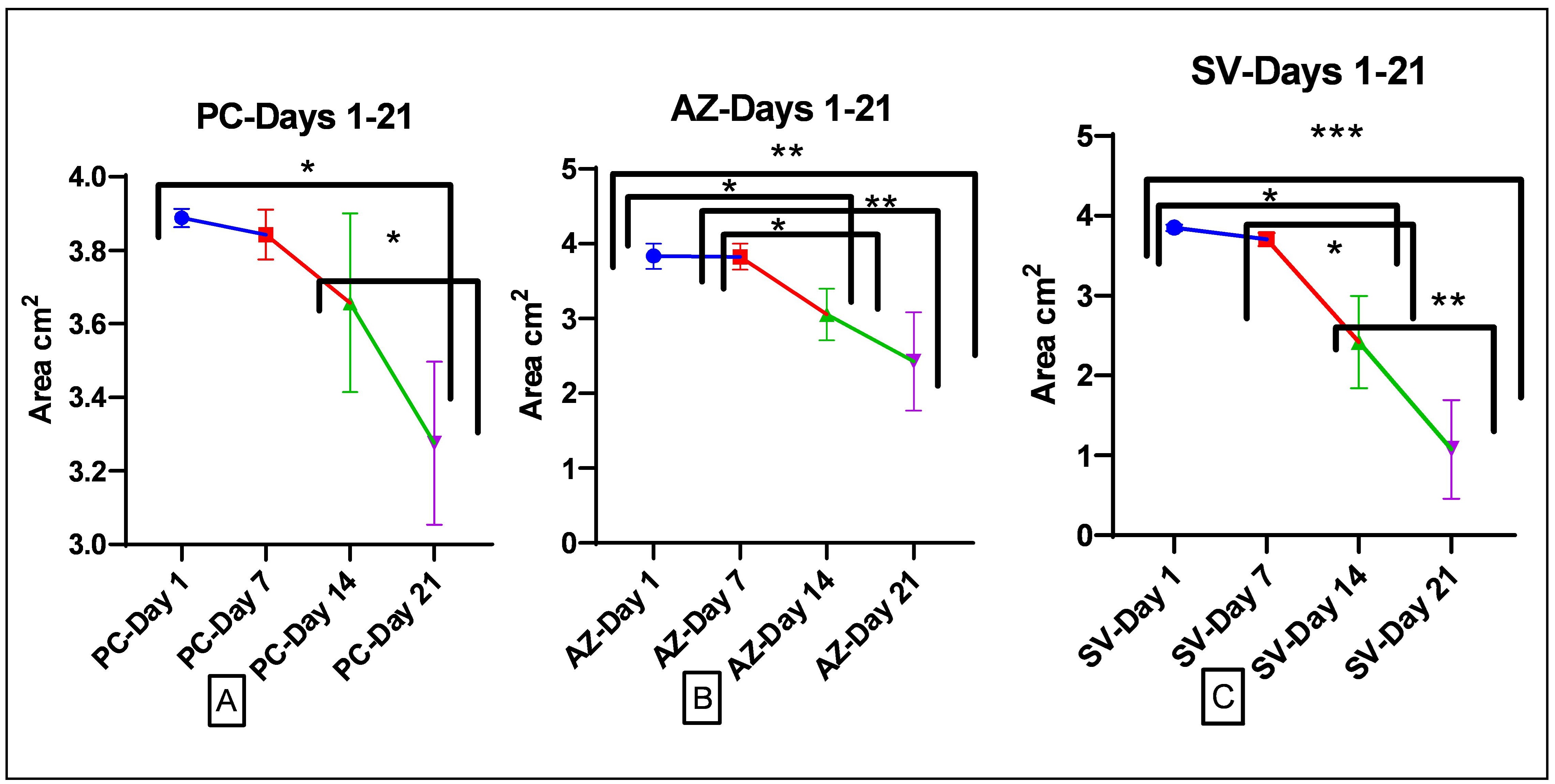
Objective: To explore the beneficial effects of azilsartan in rat models of burn wounds. Methods: Forty male rats were divided into four groups: The negative control group (NC) of 4 animals was used as a control. Positive control group (PC), azilsartan-treated group (AZ), and silver-sulfadiazine group (SV). Each group consisted of twelve rats with burn injuries, subdivided into three subgroups each of four (euthanized on days 7, 14, and 21 post-burn injury induction). Results: The levels of RBC, platelets, and HGB in the treated groups did not vary significantly. The AZ group had significantly greater WBC levels, while the AZ and SV groups had significantly higher lymphocyte levels than the PC group. After 7 days of treatment, both the PC and AZ groups showed a significant improvement in lesions and burn area, but the SV group showed no significant improvement. The improvement was considerable after 14 days of treatment in the SV-treated group and the AZ group, with no meaningful changes evident after 7 days of treatment in either of the indicated groups. When compared to the initial day of induction, no significant reduction was found in the PC group after 7 and 14 days of treatment. After 21 days of induction, the control group showed a considerable reduction in lesion and burn area. On the last day of treatment, however, the AZ and SV groups showed a more dramatic decline. Conclusions: Azilsartan heals the burnt area effectively, which could be related to limiting the local effects of Ag II, and deserves to be evaluated in a clinical environment.
Downloads
References
Fountain JH, Kaur J, Lappin SL. Physiology, Renin Angiotensin System. In: StatPearls. Treasure Island (FL): StatPearls Publishing; 2023. PMID: 29261862.
Volkan G, Abdulsamed K, Emin Ş. Role of the Renin-Angiotensin-Aldosterone System in Various Disease Processes: An Overview. In: Samy IM, (editor), Renin-Angiotensin Aldosterone System. Rijeka: IntechOpen; 2021. p. Ch. 3.
Nehme A, Cerutti C, Dhaouadi N, Gustin MP, Courand P-Y, Zibara K, et al. Atlas of tissue renin-angiotensin-aldosterone system in human: a transcriptomic meta-analysis. Sci Rep. 2015;5(1):10035. doi: 10.1038/srep10035.
Paul M, Poyan Mehr A, Kreutz R. Physiology of local renin-angiotensin systems. Physiol Rev. 2006;86(3):747-803. doi: 10.1152/physrev.00036.2005.
Akershoek JJ, Vlig M, Brouwer K, Talhout W, Beelen RHJ, Middelkoop E, et al. The presence of tissue renin-angiotensin system components in human burn wounds and scars. Burns Open. 2018;2(3):114-121. doi: 10.1016/j.burnso.2018.06.001.
Silva IMS, Assersen KB, Willadsen NN, Jepsen J, Artuc M, Steckelings UM. The role of the renin-angiotensin system in skin physiology and pathophysiology. Exp Dermatol. 2020;29(9):891-901. doi: 10.1111/exd.14159.
Hedayatyanfard K, Khalili A, Karim H, Nooraei S, Khosravi E, Haddadi NS, et al. Potential use of angiotensin receptor blockers in skin pathologies. Iran J Basic Med Sci. 2023;26(7):732-737. doi: 10.22038/IJBMS.2023.66563.14606.
Hayden MR, Sowers KM, Pulakat L, Joginpally T, Krueger B, Whaley-Connell A, et al. Possible mechanisms of local tissue renin-angiotensin system activation in the cardiorenal metabolic syndrome and type 2 diabetes mellitus. Cardiorenal Med. 2011;1(3):193-210. doi: 10.1159/000329926.
Santos RAS, Oudit GY, Verano-Braga T, Canta G, Steckelings UM, Bader M. The renin-angiotensin system: going beyond the classical paradigms. Am J Physiol Heart Circ Physiol. 2019;316(5):H958-H970. doi: 10.1152/ajpheart.00723.2018.
Jeschke MG, van Baar ME, Choudhry MA, Chung KK, Gibran NS, Logsetty S. Burn injury. Nat Rev Dis Primers. 2020;6(1):11. doi: 10.1038/s41572-020-0145-5.
Salhi N, El Guourrami O, Rouas L, Moussaid S, Moutawalli A, Benkhouili FZ, et al. Evaluation of the wound healing potential of Cynara humilis extracts in the treatment of skin burns. Evid Based Complement Alternat Med. 2023;2023:5855948. doi: 10.1155/2023/5855948.
Kim DY, Han YS, Kim SR, Chun BK, Park JH. Effects of a topical angiotensin-converting enzyme inhibitor and a selective COX-2 inhibitor on the prevention of hypertrophic scarring in the skin of a rabbit ear. Wounds. 2012;24(12):356-364. PMID: 25876220.
Akershoek JJJ, Brouwer KM, Vlig M, Boekema BKHL, Beelen RHJ, Middelkoop E, et al. Early intervention by Captopril does not improve wound healing of partial thickness burn wounds in a rat model. Burns. 2018;44(2):429-435. doi: 10.1016/j.burns.2017.08.008.
Yahata Y, Shirakata Y, Tokumaru S, Yang L, Dai X, Tohyama M, et al. A novel function of angiotensin II in skin wound healing. Induction of fibroblast and keratinocyte migration by angiotensin II via heparin-binding epidermal growth factor (EGF)-like growth factor-mediated EGF receptor transactivation. J Biol Chem. 2006;281(19):13209-13216. doi: 10.1074/jbc.M509771200.
Abadir P, Hosseini S, Faghih M, Ansari A, Lay F, Smith B, et al. Topical reformulation of valsartan for treatment of chronic diabetic wounds. J Invest Dermatol. 2018;138(2):434-443. doi: 10.1016/j.jid.2017.09.030.
Kamber M, Papalazarou V, Rouni G, Papageorgopoulou E, Papalois A, Kostourou V. Angiotensin II inhibitor facilitates epidermal wound regeneration in diabetic mice. Front Physiol. 2015;6:170. doi: 10.3389/fphys.2015.00170.
Hedayatyanfard K, Ziai SA, Niazi F, Habibi I, Habibi B, Moravvej H. Losartan ointment relieves hypertrophic scars and keloid: A pilot study. Wound Repair Regen. 2018;26(4):340-343. doi: 10.1111/wrr.12648.
Hu YY, Fang QQ, Wang XF, Zhao WY, Zheng B, Zhang DD, et al. Angiotensin-converting enzyme inhibitor and angiotensin II type 1 receptor blocker: Potential agents to reduce post-surgical scar formation in humans. Basic Clin Pharmacol Toxicol. 2020;127(6):488-494. doi: 10.1111/bcpt.13458.
Prajapati P, Zehra A, Maurya P, Singh J, Kumar A, Kushwaha S. Evaluation of azilsartan transdermal hydrogel in volumetric muscle loss injury in a rat model. J Pharm Negative Results. 2022;13:2517-2522. doi: 10.47750/pnr.2022.13.S05.389.
Rodgers KE, Dizerega GS. Contribution of the local RAS to hematopoietic function: A novel therapeutic target. Front Endocrinol (Lausanne). 2013;4:157. doi: 10.3389/fendo.2013.00157.
Carroll MA, Kang Y, Chander PN, Stier CT. Azilsartan is associated with increased circulating angiotensin-(1-7) levels and reduced renovascular 20-HETE levels. Am J Hypertens. 2015;28(5):664-671. doi: 10.1093/ajh/hpu201.
Heringer-Walther S, Eckert K, Schumacher SM, Uharek L, Wulf-Goldenberg A, Gembardt F, et al. Angiotensin-(1-7) stimulates hematopoietic progenitor cells in vitro and in vivo. Haematologica. 2009;94(6):857-860. doi: 10.3324/haematol.2008.000034.
Marathias KP, Agroyannis B, Mavromoustakos T, Matsoukas J, Vlahakos DV. Hematocrit-lowering effect following inactivation of renin-angiotensin system with angiotensin converting enzyme inhibitors and angiotensin receptor blockers. Curr Top Med Chem. 2004;4(4):483-486. doi: 10.2174/1568026043451311.
Cooper ME. The role of the renin-angiotensin-aldosterone system in diabetes and its vascular complications. Am J Hypertens. 2004;17(11 Pt 2):16S-20S; doi: 10.1016/j.amjhyper.2004.08.004.
Hao SY, Ren M, Yang C, Lin DZ, Chen LH, Zhu P, et al. Activation of skin renin-angiotensin system in diabetic rats. Endocrine. 2011;39(3):242-250. doi: 10.1007/s12020-010-9428-z.
Yevdokimova N, Podpryatov S. The up-regulation of angiotensin II receptor type 1 and connective tissue growth factor are involved in high-glucose-induced fibronectin production by cultured human dermal fibroblasts. J Dermatol Sci. 2007;47(2):127-139. doi: 10.1016/j.jdermsci.2007.02.009.
Faghih M, Hosseini SM, Smith B, Ansari AM, Lay F, Ahmed AK, et al. Knockout of angiotensin AT2 receptors accelerates healing but impairs quality. Aging (Albany NY). 2015;7(12):1185-1197. doi: 10.18632/aging.100868.
Rodgers K, Xiong S, Felix J, Roda N, Espinoza T, Maldonado S, et al. Development of angiotensin (1-7) as an agent to accelerate dermal repair. Wound Repair Regen. 2001;9(3):238-247. doi: 10.1046/j.1524-475x.2001.00238.x.
Downloads
Published
How to Cite
Issue
Section
License
Copyright (c) 2024 Al-Rafidain Journal of Medical Sciences ( ISSN 2789-3219 )

This work is licensed under a Creative Commons Attribution-NonCommercial-ShareAlike 4.0 International License.
Published by Al-Rafidain University College. This is an open access journal issued under the CC BY-NC-SA 4.0 license (https://creativecommons.org/licenses/by-nc-sa/4.0/).


