Investigating the Effect of EX-527 as SIRT1 Inhibitor in Breast Cancer Cell Line
DOI:
https://doi.org/10.54133/ajms.v7i1(Special).926Keywords:
Breast cancer, EX-527, MCF-7, SIRT1 inhibitorAbstract
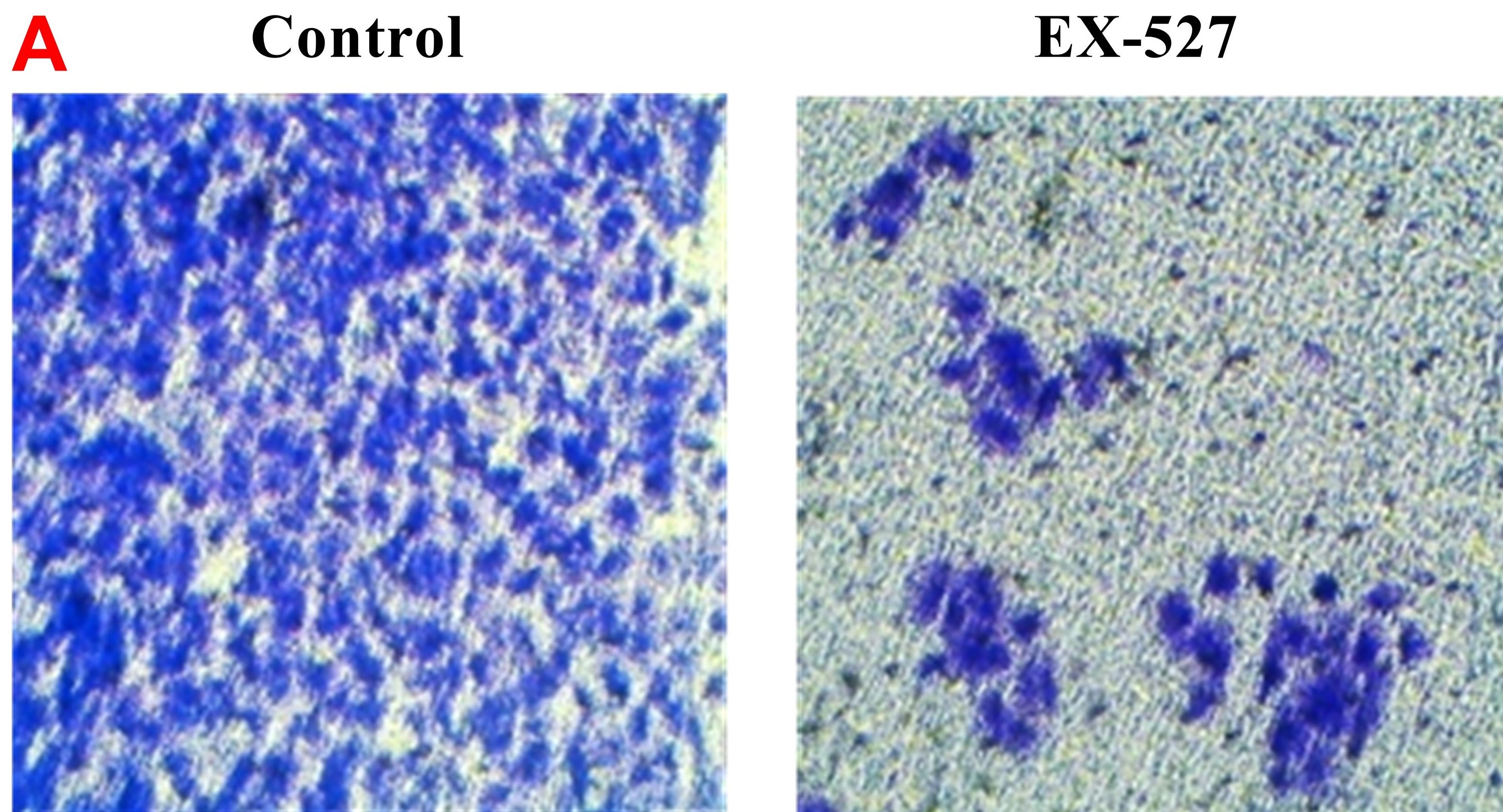
Background: Breast cancer is one of the most common malignant and metastatic tumors worldwide that cause death in women. Therefore, preventing the growth and metastasis of cancerous cells is essential for enhancing the prognosis and efficacy of treatment for breast cancer. Sirtuin 1 (SIRT1) is a nicotinamide-adenine dinucleotide (NAD+)-dependent deacetylase that has been linked to a number of biological processes, including genomic stability, cell cycle, cell survival and cancer metastasis. EX-527 is a selective and potent SIRT1 inhibitor. Recent studies have revealed that SIRT1 has an oncogenic role in breast cancer. Objective: To evaluate the effect of EX-527 on the MCF-7 breast cancer cell line. Methods: MCF-7 was cultured in complete DMEM and treated with and without EX-527. Cell viability of the breast cancer cell line was evaluated by MTT assay and apoptosis by Annexin V/PI staining. Migration and invasion of breast cancer cells were determined by wound healing and transwell invasion assays, respectively. Results: Results revealed that EX-527 at a concentration of 25.30 µM was associated with a significant anti-proliferative effect and induction of apoptosis (98.3%) in breast cancer cells. Treatment with EX-527 was also associated with significant suppression of migration and invasion of MCF-7. Conclusions: The current investigation showed that inhibition of SIRT1 by EX-527 inhibits proliferation, migration, invasion and apoptosis of human breast cancer cells.
Downloads
References
Bray F, Ferlay J, Soerjomataram I, Siegel RL, Torre LA, Jemal A. Global cancer statistics 2018: GLOBOCAN estimates of incidence and mortality worldwide for 36 cancers in 185 countries. Cancer J Clin. 2018;68(6):394-424. doi: 10.3322/caac.21492.
Torre LA, Bray F, Siegel RL, Ferlay J, Lortet-Tieulent J, Jemal A. Global cancer statistics, 2012. CA Cancer J Clin. 2015;65(2):87-108. doi: 10.3322/caac.21262.
DeSantis CE, Ma J, Goding Sauer A, Newman LA, Jemal A. Breast cancer statistics, 2017, racial disparity in mortality by state. CA Cancer J Clin. 2017;67(6):439-448. doi: 10.3322/caac.21412.
Jiang K, Song X, Yang L, Li L, Wan Z, Sun X, et al. Enhanced antitumor and anti-metastasis efficacy against aggressive breast cancer with a fibronectin-targeting liposomal doxorubicin. J Control Release. 2018;271:21-30. doi: 10.1016/j.jconrel.2017.12.026.
Medeiros B, Allan AL. Molecular mechanisms of breast cancer metastasis to the lung: Clinical and experimental perspectives. Int J Mol Sci. 2019;20(9):2272. doi: 10.3390/ijms20092272.
Theodossiou TA, Ali M, Grigalavicius M, Grallert B, Dillard P, Schink KO, et al. Simultaneous defeat of MCF7 and MDA-MB-231 resistances by a hypericin PDT-tamoxifen hybrid therapy. NPJ Breast Cancer. 2019;5:13. doi: 10.1038/s41523-019-0108-8.
Grubczak K, Kretowska-Grunwald A, Groth D, Poplawska I, Eljaszewicz A, Bolkun L, et al. Differential response of MDA-MB-231 and MCF-7 breast cancer cells to in vitro inhibition with CTLA-4 and PD-1 through cancer-immune cells modified interactions. Cells. 2021;10(8):2044. doi: 10.3390/cells10082044.
Huang KS, Wang YT, Byadgi O, Huang TY, Tai MH, Shaw JF, et al. Screening of specific and common pathways in breast cancer cell lines MCF-7 and MDA-MB-231 treated with chlorophyllides composites. Molecules. 2022;27(12):3950. doi: 10.3390/molecules27123950.
Sauve AA, Wolberger C, Schramm VL, Boeke JD. The biochemistry of sirtuins. Annu Rev Biochem. 2006;75:435-465. doi: 10.1146/annurev.biochem.74.082803.133500.
Michan S, Sinclair D. Sirtuins in mammals: insights into their biological function. Biochem J. 2007;404(1):1-13. doi: 10.1042/BJ20070140.
Karbasforooshan H, Roohbakhsh A, Karimi G. SIRT1 and microRNAs: The role in breast, lung and prostate cancers. Exp Cell Res. 2018;367(1):1-6. doi: 10.1016/j.yexcr.2018.03.023.
Liu T, Liu PY, Marshall GM. The critical role of the class III histone deacetylase SIRT1 in cancer. Cancer Res. 2009;69(5):1702-1705. doi: 10.1158/0008-5472.CAN-08-3365.
Antoniali G, Marcuzzi F, Casarano E, Tell G. Cadmium treatment suppresses DNA polymerase δ catalytic subunit gene expression by acting on the p53 and Sp1 regulatory axis. DNA Repair. 2015;35:90-105. doi: 10.1016/j.dnarep.2015.08.007.
Luo J, Nikolaev AY, Imai S, Chen D, Su F, Shiloh A, et al. Negative control of p53 by Sir2alpha promotes cell survival under stress. Cell. 2001;107(2):137-148. doi: 10.1016/s0092-8674(01)00524-4.
Peck B, Chen CY, Ho KK, Di Fruscia P, Myatt SS, Coombes RC, et al. SIRT inhibitors induce cell death and p53 acetylation through targeting both SIRT1 and SIRT2. Mol Cancer Ther. 2010;9(4):844-855. doi: 10.1158/1535-7163.MCT-09-0971.
Solomon JM, Pasupuleti R, Xu L, McDonagh T, Curtis R, DiStefano PS, et al. Inhibition of SIRT1 catalytic activity increases p53 acetylation but does not alter cell survival following DNA damage. Mol Cell Biol. 2006;26(1):28-38. doi: 10.1128/MCB.26.1.28-38.2006.
Asaka R, Miyamoto T, Yamada Y, Ando H, Mvunta DH, Kobara H, et al. Sirtuin 1 promotes the growth and cisplatin resistance of endometrial carcinoma cells: a novel therapeutic target. Lab Invest. 2015;95(12):1363-1373. doi: 10.1038/labinvest.2015.119.
Chen G, Zhang B, Xu H, Sun Y, Shi Y, et al . Suppression of Sirt1 sensitizes lung cancer cells to WEE1 inhibitor MK-1775-induced DNA damage and apoptosis. Oncogene. 2017;36(50):6863-6872. doi: 10.1038/onc.2017.297.
Chen MC, Yu CH, Wang SW, Pu HF, Kan SF, Lin LC, et al. Anti-proliferative effects of evodiamine on human thyroid cancer cell line ARO. J Cell Biochem. 2010;110(6):1495-503. doi: 10.1002/jcb.22716.
Kamiloglu S, Sari G, Ozdal T, Capanoglu E. Guidelines for cell viability assays. Food Front. 2020;1(3):332-349. doi: 10.1002/fft2.44.
Salman RF, Al-Sudani BT, Mshimesh BA. Protective action of SIRT1 activator aptamer in human skin cell line. J Popl Ther Clin Pharmacol. 2023;30(5):342-359. doi: 10.47750/jptcp.2023.30.05.035.
Crowley LC, Marfell BJ, Scott AP, Waterhouse NJ. Quantitation of apoptosis and necrosis by annexin V binding, propidium iodide uptake, and flow cytometry. Cold Spring Harb Protoc. 2016;2016(11). doi: 10.1101/pdb.prot087288.
Kozlova N, Samoylenko A, Drobot L, Kietzmann T. Urokinase is a negative modulator of Egf-dependent proliferation and motility in the two breast cancer cell lines MCF-7 and MDA-MB-231. Mol Carcinog. 2016;55(2):170-181. doi: 10.1002/mc.22267.
Al-khfajy WS, Arif IS, Al-Sudani BT. Synergistic effect of obeticholic acid and fasting-mimicking on proliferative, migration, and survival signaling in prostate cancer. Pharmacia. 2022;69(2):579-587. doi: 10.3897/pharmacia.69.e81452.
Brackstone M, Townson JL, Chambers AF. Tumour dormancy in breast cancer: an update. Breast Cancer Res. 2007;9(3):208. doi: 10.1186/bcr1677.
Nounou MI, ElAmrawy F, Ahmed N, Abdelraouf K, Goda S, Syed-Sha-Qhattal H. Breast cancer: Conventional diagnosis and treatment modalities and recent patents and technologies. Breast Cancer (Auckl). 2015 ;9(Suppl 2):17-34. doi: 10.4137/BCBCR.S29420.
Arnedos M, Bihan C, Delaloge S, Andre F. Triple-negative breast cancer: are we making headway at least? Ther Adv Med Oncol. 2012;4(4):195-210. doi: 10.1177/1758834012444711.
Wang C, Yang W, Dong F, Guo Y, Tan J, Ruan S, et al. The prognostic role of Sirt1 expression in solid malignancies: a meta-analysis. Oncotarget. 2017;8(39):66343-66351. doi: 10.18632/oncotarget.
Nin V, Escande C, Chini CC, Giri S, Camacho-Pereira J, Matalonga J, et al. Role of deleted in breast cancer 1 (DBC1) protein in SIRT1 deacetylase activation induced by protein kinase A and AMP-activated protein kinase. J Biol Chem. 2012;287(28):23489-23501. doi: 10.1074/jbc.M112.365874.
Jin X, Wei Y, Xu F, Zhao M, Dai K, Shen R, et al. SIRT1 promotes formation of breast cancer through modulating Akt activity. J Cancer. 2018;9(11):2012-2023. doi: 10.7150/jca.24275.
Wawruszak A, Okon E, Telejko I, Czerwonka A, Luszczki J. Additive pharmacological interaction between sirtuin inhibitor cambinol and paclitaxel in MCF7 luminal and MDA-MB-231 triple-negative breast cancer cells. Pharmacol Rep. 2022;74(5):1011-1024. doi: 10.1007/s43440-022-00393-w.
Zou Q, Tang Q, Pan Y, Wang X, Dong X, Liang Z, et al. MicroRNA-22 inhibits cell growth and metastasis in breast cancer via targeting of SIRT1. Exp Ther Med. 2017;14(2):1009-1016. doi: 10.3892/etm.2017.4590.
Abdolvahabi Z, Nourbakhsh M, Hosseinkhani S, Hesari Z, Alipour M, Jafarzadeh M, et al. MicroRNA-590-3P suppresses cell survival and triggers breast cancer cell apoptosis via targeting sirtuin-1 and deacetylation of p53. J Cell Biochem. 2019;120(6):9356-9368. doi: 10.1002/jcb.28211.
Lin Z, Fang D. The roles of SIRT1 in cancer. Genes Cancer. 2013;4(3-4):97-104. doi: 10.1177/1947601912475079.
Zhang X, Li Y, Wang D, Wei X. miR-22 suppresses tumorigenesis and improves radiosensitivity of breast cancer cells by targeting Sirt1. Biol Res. 2017;50(1):27. doi: 10.1186/s40659-017-0133-8.
Kalle AM, Mallika A, Badiger J, Alinakhi, Talukdar P, Sachchidanand. Inhibition of SIRT1 by a small molecule induces apoptosis in breast cancer cells. Biochem Biophys Res Commun. 2010;401(1):13-19. doi: 10.1016/j.bbrc.2010.08.118.
Peck B, Chen CY, Ho KK, Di Fruscia P, Myatt SS, Coombes RC, et al. SIRT inhibitors induce cell death and p53 acetylation through targeting both SIRT1 and SIRT2. Mol Cancer Ther. 2010;9(4):844-855. doi: 10.1158/1535-7163.MCT-09-0971.
Edelstein ML, Abedi MR, Wixon J. Gene therapy clinical trials worldwide to 2007--an update. J Gene Med. 2007;9(10):833-842. doi: 10.1002/jgm.1100.
Xu Y, Qin Q, Chen R, Wei C, Mo Q. SIRT1 promotes proliferation, migration, and invasion of breast cancer cell line MCF-7 by upregulating DNA polymerase delta1 (POLD1). Biochem Biophys Res Commun. 2018;502(3):351-357. doi: 10.1016/j.bbrc.2018.05.164.
Kim HW, Kim SA, Ahn SG. Sirtuin inhibitors, EX527 and AGK2, suppress cell migration by inhibiting HSF1 protein stability. Oncol Rep. 2016;35(1):235-242. doi: 10.3892/or.2015.4381.
Li L, Xie X, Luo J, Liu M, Xi S, Guo J, et al. Targeted expression of miR-34a using the T-VISA system suppresses breast cancer cell growth and invasion. Mol Ther. 2012;20:2326–2334. doi: 10.1002/anie.202218872.
Downloads
Published
How to Cite
Issue
Section
License
Copyright (c) 2024 Al-Rafidain Journal of Medical Sciences ( ISSN 2789-3219 )

This work is licensed under a Creative Commons Attribution-NonCommercial-ShareAlike 4.0 International License.
Published by Al-Rafidain University College. This is an open access journal issued under the CC BY-NC-SA 4.0 license (https://creativecommons.org/licenses/by-nc-sa/4.0/).


